Cell line - MCF10A
MCF10A is a non-transformed epithelial cell line. Images for this cell line have been generated with a BioTek Cytation5 under environmental control (37 ℃ and 5% CO2) in one field of view. The cells were maintained in a BioTek Biospa8 automated incubator (37 ℃ and 5% CO2) during the interval period of imaging. All images have the nuclear reporter H2b-mRuby.
The cell line was seeded onto 12-well plates (Corning Falcon #353043). The seeding density was 2-3×10⁴ cells/well. Cells were incubated for 16 hours at 37 ℃ and 5% CO2, then the medium was changed to remove dead cells and further incubated for 2 hours. Images were acquired at 4X, 10X, and 20X magnifications with the RFP channel (531/593) to visualize red fluorescent protein, mRuby, as well as the phase contrast channel with 10-15 minute intervals for three days.
The MCF10A cells H2b-mRuby were cultured in DMEM/F12 (Gibco #11330-032) supplemented with 5% horse serum (Gibco #16050122), 20 ng/mL EGF (PeproTech #AF100-15), 0.5 mg/mL Hydrocortisone (Sigma #H0888), 100 ng/mL cholera toxin (Sigma #C8052), and 10 µg/mL insulin (Sigma #I-1882) containing hygromycin (Millipore Sigma #H3274-100MG) (1.5 µg/mL) and puromycin (STEMCELL #73342) (0.1 µg/mL). HEK293T cells expressing H2b-mRuby were cultured in DMEM (Gibco #10313021) supplemented with 10% FBS (V/V) (Gibco #10082139), 2mM L-glutamine (Corning #25-005-CI) containing 0.5 µg/mL puromycin (STEMCELL #73342). For trypsinization, 0.05% trypsin, 0.53 mM EDTA (Corning #25-052-CI) was used.
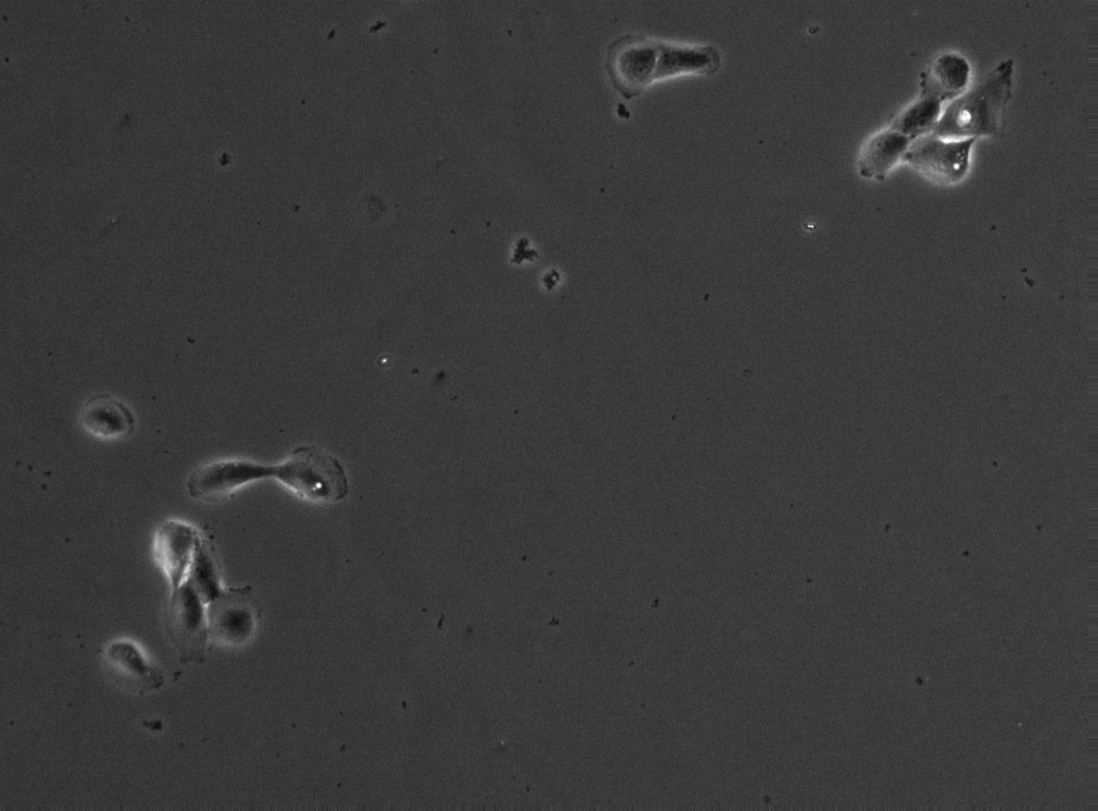
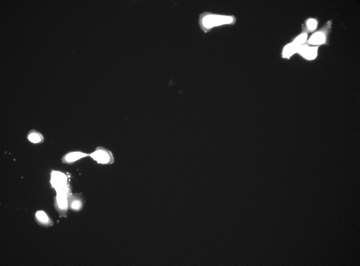
FLD_3
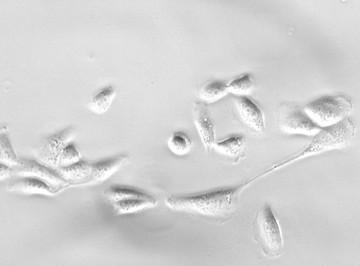
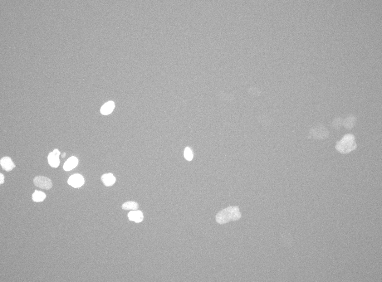
FLD_113
Example sequences from the MCF10A dataset. Left: phase contrast. Right: nuclear fluorescence (RFP).
Images
10× magnification
Annotation
Videos
10× magnification
FLD_3
GAP 1: pc | rfp | annotation
GAP 2: pc | rfp | annotation
GAP 4: pc | rfp | annotation
GAP 8: pc | rfp | annotation
FLD_4
GAP 1: pc | rfp | annotation
GAP 2: pc | rfp | annotation
GAP 4: pc | rfp | annotation
GAP 8: pc | rfp | annotation
FLD_5
GAP 1: pc | rfp | annotation
GAP 2: pc | rfp | annotation
GAP 4: pc | rfp | annotation
GAP 8: pc | rfp | annotation
FLD_6
GAP 1: pc | rfp | annotation
GAP 2: pc | rfp | annotation
GAP 4: pc | rfp | annotation
GAP 8: pc | rfp | annotation
FLD_7
GAP 1: pc | rfp | annotation
GAP 2: pc | rfp | annotation
GAP 4: pc | rfp | annotation
GAP 8: pc | rfp | annotation
FLD_8
GAP 1: pc | rfp | annotation
GAP 2: pc | rfp | annotation
GAP 4: pc | rfp | annotation
GAP 8: pc | rfp | annotation
FLD_9
GAP 1: pc | rfp | annotation
GAP 2: pc | rfp | annotation
GAP 4: pc | rfp | annotation
GAP 8: pc | rfp | annotation
FLD_10
GAP 1: pc | rfp | annotation
GAP 2: pc | rfp | annotation
GAP 4: pc | rfp | annotation
GAP 8: pc | rfp | annotation
FLD_11
GAP 1: pc | rfp | annotation
GAP 2: pc | rfp | annotation
GAP 4: pc | rfp | annotation
GAP 8: pc | rfp | annotation
20× magnification
FLD_102
GAP 1: pc | rfp | annotation
GAP 2: pc | rfp | annotation
GAP 4: pc | rfp | annotation
GAP 8: pc | rfp | annotation
FLD_103
GAP 1: pc | rfp | annotation
GAP 2: pc | rfp | annotation
GAP 4: pc | rfp | annotation
GAP 8: pc | rfp | annotation
FLD_104
GAP 1: pc | rfp | annotation
GAP 2: pc | rfp | annotation
GAP 4: pc | rfp | annotation
GAP 8: pc | rfp | annotation
FLD_106
GAP 1: pc | rfp | annotation
GAP 2: pc | rfp | annotation
GAP 4: pc | rfp | annotation
GAP 8: pc | rfp | annotation
FLD_107
GAP 1: pc | rfp | annotation
GAP 2: pc | rfp | annotation
GAP 4: pc | rfp | annotation
GAP 8: pc | rfp | annotation
FLD_108
GAP 1: pc | rfp | annotation
GAP 2: pc | rfp | annotation
GAP 4: pc | rfp | annotation
GAP 8: pc | rfp | annotation
FLD_110
GAP 1: pc | rfp | annotation
GAP 2: pc | rfp | annotation
GAP 4: pc | rfp | annotation
GAP 8: pc | rfp | annotation
FLD_111
GAP 1: pc | rfp | annotation
GAP 2: pc | rfp | annotation
GAP 4: pc | rfp | annotation
GAP 8: pc | rfp | annotation
FLD_113
GAP 1: pc | rfp | annotation
GAP 2: pc | rfp | annotation
GAP 4: pc | rfp | annotation
GAP 8: pc | rfp | annotation