Cell line - U87
U87 is a human brain cancer cell line derived from malignant gliomas. Images have been generated with BioTek Cytation5 under environmental control (37 ℃ and 5% CO2) in one field of view. The cells were maintained in a BioTek Biospa8 automated incubator (37 ℃ and 5% CO2) during the interval period of imaging. Cells had the nuclear reporter H2b-mRuby.
Cell lines were seeded onto 12 well plates (Corning Falcon #353043). The seeding density were 1.5×10⁴ cells/well. Cells were incubated for 16 hours at 37 ℃ and 5% CO2, then the medium was changed to remove the dead cells and further incubated for 2 hours. Images were acquired at 10X and 20X magnifications with RFP channel (531/593) to visualize red fluorescent protein, mRuby as well as Phase contrast channel with 15 minutes interval for three days.
U87 cells expressing H2b-mRuby were cultured in DMEM (Gibco #10313021) supplemented with 10% FBS (V/V) (Gibco #10082139), 2mM L-glutamine (Corning #25-005-CI) containing 0.2 µg/mL puromycin (STEMCELL #73342). For trypsinization 0.05% Trypsin-EDTA (Gibco, #25200-072) was used.
Generation of U87 Cell Line Expressing H2b-mRuby
Lentivirus vector is used to deliver pLenti-puro-H2B-mRuby plasmid to the cells. Co-transfection of HEK293T Cells to Make Third Generation Lentivirus 5 cells were seeded onto a T75 flask (Greiner #658175) the day before transfection and incubated for 22 hours. The media was changed to serum free media (DMEM, 10% FBS) and incubated for two hours. Co-transfection was performed using 6000 ng of the packaging plasmid, pPAX (Addgene, #12260), 3000 ng of envelop plasmid, pCMV-VSV-G (Addgene #8454) and 15000 ng of transfer plasmid with lipofectamine 2000 (Thermo-Fisher # 11668027) using manufacturer’s protocol. The media changed with the full growth media (DMEM, 10% FBS and 2 mM L glutamine) after 6 hours post transfection. First lentiviral supernatant was collected after 48 hours of post transfection and warm full growth media was added to the T75 flask. The lentiviral supernatant was again collected after 72 hours post transfection. The combined supernatant was then centrifuged at 1000xG for 5 minutes to pellet any cell debris. The supernatant was concentrated using Amicon Ultra-15 100 kDa centrifugal filter (Millipore # UFC910008), aliquoted and preserved at -80.
Lentiviral Transduction of Cells 1 cell suspension was transduced in full growth media with 150 L of the concentrated lentiviral supernatant in a 6 well plate (Corning Falcon #353046). 48-hour post-transduction the expression was verified by fluorescence imaging. All the cell lines were selected with selected with puromycin (2 g/mL, STEMCELL #73342) for a week until all negative control cells were dead. Stable H2B-mRuby expressing cells were maintained in full growth medium with 0.2 g/mL puromycin.
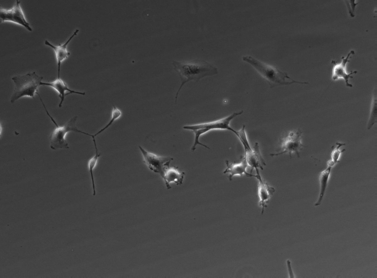
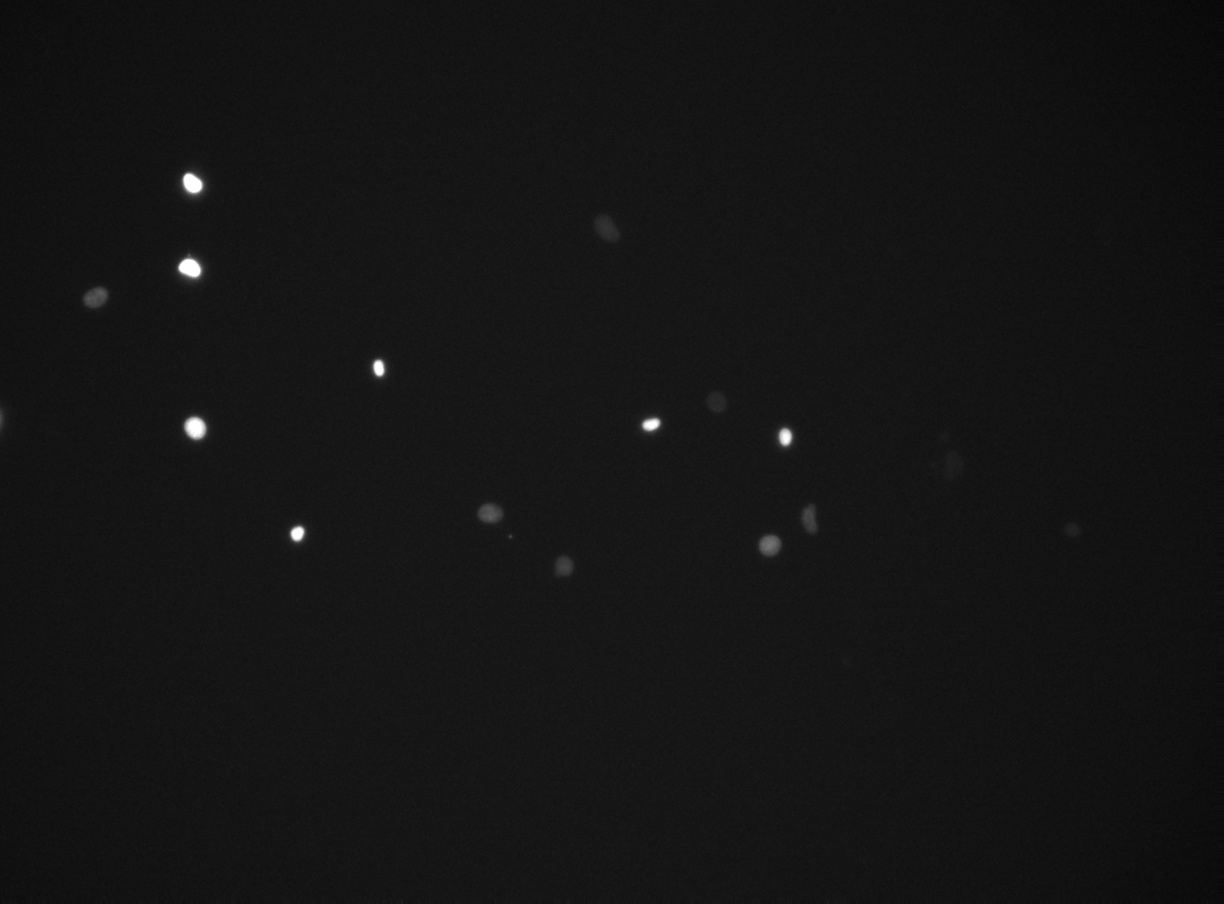
FLD_12
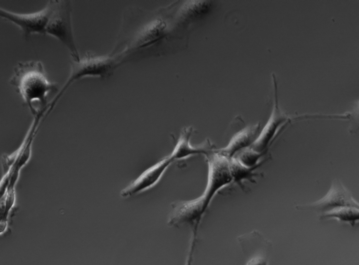
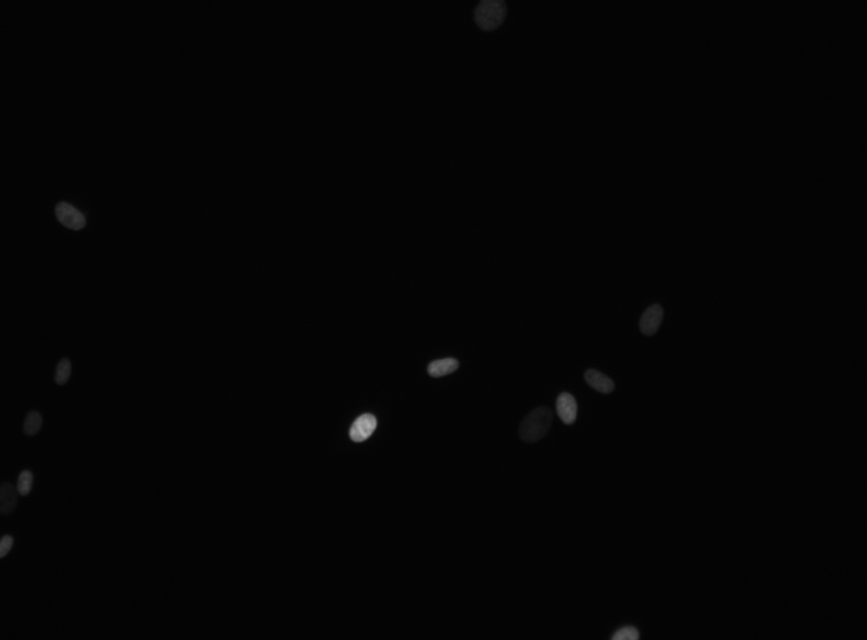
FLD_1
Example sequences from the U87 dataset. Left: phase contrast. Right: nuclear fluorescence (RFP).
Images
Annotation
Videos
10× magnification
FLD_12
GAP 1: pc | rfp | annotation
GAP 2: pc | rfp | annotation
GAP 4: pc | rfp | annotation
GAP 8: pc | rfp | annotation
FLD_13
GAP 1: pc | rfp | annotation
GAP 2: pc | rfp | annotation
GAP 4: pc | rfp | annotation
GAP 8: pc | rfp | annotation
FLD_17
GAP 1: pc | rfp | annotation
GAP 2: pc | rfp | annotation
GAP 4: pc | rfp | annotation
GAP 8: pc | rfp | annotation
FLD_19
GAP 1: pc | rfp | annotation
GAP 2: pc | rfp | annotation
GAP 4: pc | rfp | annotation
GAP 8: pc | rfp | annotation
20× magnification
FLD_1
GAP 1: pc | rfp | annotation
GAP 2: pc | rfp | annotation
GAP 4: pc | rfp | annotation
GAP 8: pc | rfp | annotation
FLD_2
GAP 1: pc | rfp | annotation
GAP 2: pc | rfp | annotation
GAP 4: pc | rfp | annotation
GAP 8: pc | rfp | annotation
FLD_3
GAP 1: pc | rfp | annotation
GAP 2: pc | rfp | annotation
GAP 4: pc | rfp | annotation
GAP 8: pc | rfp | annotation
FLD_4
GAP 1: pc | rfp | annotation
GAP 2: pc | rfp | annotation
GAP 4: pc | rfp | annotation
GAP 8: pc | rfp | annotation